CRIME Greatest Violations of Nuremberg Code in History – Catherine Austin Fitts/Greg Hunter

Dozdoats

On TB every waking moment
Greatest Violations of Nuremberg Code in History – Catherine Austin Fitts | Greg Hunter’s USAWatchdog

RT 34:35

Greatest Violations of Nuremberg Code in History – Catherine Austin Fitts
By Greg Hunter On April 17, 2021 In Political Analysis 60 Comments


By Greg Hunter’s USAWatchdog.com
(Saturday Night Post 4.17.2021)

Investment advisor and former Assistant Secretary of Housing Catherine Austin Fitts contends CV19 and the vaccines to cure it are more about control than depopulation. Fitts explains, “I think the bankers are trying to chip us. Moderna describes their injection, gene therapy as an ‘operating system.’ I agree with them. I think they are trying to download an operating system into our bodies. I don’t think it was an accident . . . the man President Trump appointed as head of ‘Operation Warp Speed’ was an expert at Brain-Machine interface. . . . Just like Bill Gates downloaded an operating system into your computer and made you update it regularly because of the threat of another virus, I think they are trying to play the same game with human bodies. It’s hard for people to fathom if they have not been following the advancements in biotech and to fathom how much money the bankers can make if they can achieve this. We just saw the Chairman of the Federal Reserve talking about the economy was getting better because the vaccination rate was going up. I think that’s code for the bank stocks are going up because we are downloading operating systems in more and more people, and our stock reflects that. We get a pop on our stock for every person we can remotely control with our operating system. . . . If you look at the deaths and adverse events, and the failure to provide true informed consent, we are talking about the greatest violations of the Nuremberg Code in history—now.”

Fitts says don’t believe the hype on the number of CV19 vaccines being given. Fitts explains, “One of the things I have seen and gotten feedback on is that the resistance is much greater than anything they are indicating in any kind of official statistics. There are also indications that the deaths and adverse events (from the vaccines) are much worse, and that has to be spreading virally. If you look at the people most resistant, including healthcare workers and nursing staff, they are seeing the adverse events, and they are seeing the deaths. So, I don’t trust the statistics. . . . The top doctors I trust essentially say this is an experiment, and it’s true. These vaccines are not approved by the FDA. These are authorized under experimental use. So, this is a trial, a human trial. The doctors I trust say we won’t know for 4, 6, 12 or 18 months what the real impact is. These are not vaccines. It is gene therapy and downloading an operating system. I would argue that they are not vaccinations, but whatever they are, if it follows the history of vaccinations, what you are going to see is a tremendous diminution of people’s immune systems and a whole world of autoimmune diseases that can be explained away by other things. I would guess that the leadership’s goal is not necessarily to depopulate, and I could be wrong, but their goal is to install an operating system. To get that done, they don’t care how many people they kill.”

In closing, Fitts says, “Naomi Wolf was giving an interview about the vaccine passports, and she said this is the end of human liberty in the west, and that’s right. If those things are allowed, along with the operating system, it is the end of liberty. We are talking about a slavery system. . . . The greatest navigation tool ever created is prayer.”

Join Greg Hunter of USAWatchdog.com as he goes One-on-One with Catherine Austin Fitts, publisher of The Solari Report.
(To Donate to USAWatchdog.com Click Here)


After the Interview:
Catherine Austin Fitts also said, “It’s against the law to lie about bio-hazards (CV19 and vaccines), and that is exactly what they are doing in my mind.

There is much free information on Solari.com, including the free report called “A Sane Person’s Guidebook to the Global Pandemic.”

You can get way more cutting edge analysis from Catherine Austin Fitts and “The Solari Report” by becoming a subscriber.
 
Writing
Heard in a recent video that in the stimulus $2.3 Billion dollars was going to Gates.
I have a low intelligence friend who brags that he got the two shots. I had told him not to get it, but he really either didn't understand or didn't believe me. He offered no input. I hang with him to help him. He's 65, poor, no car and just got out of alcohol detox. He is sorta pitiful. I am waiting to see if he gets real sick in a year's time.

I guess he's my bird in the coal mine. My cousins wife I doubt will tell me anything if she gets sick. Writing this from my phone. My PC had crashed for a month,but I finally got it fixed. In the tub now so hope I don't drop the phone. Holding it tight.
 

Seeker22

Has No Life - Lives on TB
Writing
I have a low intelligence friend who brags that he got the two shots. I had told him not to get it, but he really either didn't understand or didn't believe me. He offered no input. I hang with him to help him. He's 65, poor, no car and just got out of alcohol detox. He is sorta pitiful. I am waiting to see if he gets real sick in a year's time.

I guess he's my bird in the coal mine. My cousins wife I doubt will tell me anything if she gets sick. Writing this from my phone. My PC had crashed for a month,but I finally got it fixed. In the tub now so hope I don't drop the phone. Holding it tight.

That's usually when the little puppy flies out of my grasp.

When are adults going to do what is best for them instead of acting like insecure teenagers and bowing to peer pressure? Sheep in a herd.
If this is what "adult" looks like- WTH did I bother to grow up? There are so many reasons not to take this so called vax.
 
Ha ha yeah. I should learn cuz 2 months ago I dropped the old phone in a bowl of water. But anyway I like CAF's. She is really on the ball. I couldn't get into the site to read the free guidebook. Said log in, but no place to register. Will try again.
 

Hfcomms

EN66iq
I like him but he has a bad habit of interrupting his guests all the time and he is supposed to be interviewing them. That being said two things stuck out. The first one is the Pentagon admitting to verification of UFO's in June which is supposed to turn things upside down and that the 'vaccine' thru the gene therapy is indeed an operating system to make people more susceptible to mind control operations.

I have heard both of these things before but Catherine buying into them is certainly interesting.
 

Milkweed Host

Veteran Member
Hunter was interrupting way too much in this excellent interview.
I was hoping that Fitts would have provided more info in the 'How' this jap will
affect people, some details. She must have a reason for her position?
 

Cardinal

Chickministrator
_______________
I like him but he has a bad habit of interrupting his guests all the time and he is supposed to be interviewing them.
He strikes me as being some alphabet combo-ADD or ADHD or something along those lines.
 

Hfcomms

EN66iq
Not going to bash him too hard. You get what you pay for and in this case it costs us nothing. He is an honest broker with his own bias as we all have and he does have some thought provoking guests.
 

Dozdoats

On TB every waking moment
I don't know if he was flummoxed by what she said, upset by other things or what was going on, but he seemed to be channeling his inner 8YO much more than usual.
 

Walrus Whisperer

Hope in chains...
Writing
I have a low intelligence friend who brags that he got the two shots. I had told him not to get it, but he really either didn't understand or didn't believe me. He offered no input. I hang with him to help him. He's 65, poor, no car and just got out of alcohol detox. He is sorta pitiful. I am waiting to see if he gets real sick in a year's time.

I guess he's my bird in the coal mine. My cousins wife I doubt will tell me anything if she gets sick. Writing this from my phone. My PC had crashed for a month,but I finally got it fixed. In the tub now so hope I don't drop the phone. Holding it tight.
So WHY in the HELL are you hanging around a low intelligence "friend"? You CANNOT help every lowlife bum. You've gotten yourself in trouble before trying to "be nice". You, Mary, may end up killed because of your propensity to gather forlorn unsuitable people around you.
STOP IT!
 
So WHY in the HELL are you hanging around a low intelligence "friend"? You CANNOT help every lowlife bum. You've gotten yourself in trouble before trying to "be nice". You, Mary, may end up killed because of your propensity to gather forlorn unsuitable people around you.
STOP IT!
Well, he is very passive and a gentle person. I wonder if I got a little bit of "liberal" in me cuz I am always nice to people and my whole life people been mean to me. He used to be a hard worker and took care of his old mom. Then he fell and broke his hip so can't work anymore. He is 66 now.
 

China Connection

TB Fanatic
Moncef Slaoui Departs Galvani Bioelectronics Board of Directors
For media and investors only
Issued: London, UK
The Board of Directors of GlaxoSmithKline plc (“GSK”), the majority shareholder of Galvani Bioelectronics (“Galvani”), today announced the termination of Moncef Slaoui as Chair of the Galvani Board of Directors, effective immediately.
The termination follows the receipt of a letter containing allegations of sexual harassment and inappropriate conduct towards an employee of GSK by Dr. Slaoui, which occurred several years ago when he was an employee of GSK. Upon receipt of the letter, the GSK Board immediately initiated an investigation with an experienced law firm to investigate the allegations. The investigation of Dr. Slaoui’s conduct substantiated the allegations and is ongoing.
Dr. Slaoui’s behaviours are wholly unacceptable. They represent an abuse of his leadership position, violate company policies, and are contrary to the strong values that define GSK’s culture. The company expects everyone at GSK to behave in accordance with its values, especially its leaders where its standards are the highest. Sexual harassment and any abuse of leadership position are strictly prohibited and will not be tolerated.
Christopher Corsico, SVP Development at GSK and a current member of the Galvani Board, has been appointed as the new Chair of the Board of Galvani. In addition, Amy Altshul, SVP Legal, R&D and Global Commercial Franchises at GSK, has also been appointed to the Board.
GSK’s leadership is firmly committed to building a safe and respectful environment for every employee. The company has established policies and resources to manage issues related to workplace safety and conduct while protecting the privacy and wellbeing of its employees. Over the last few years, it has worked hard to prioritise and enhance efforts to ensure that all employees feel respected and included.
About GSK
GSK is a science-led global healthcare company with a special purpose: to help people do more, feel better, live longer. For further information please visit www.gsk.com/about-us.
About Galvani Bioelectronics

Galvani Bioelectronics is a medical research company dedicated to the development of bioelectronic medicines to treat chronic diseases. Formed through a partnership between two global healthcare companies, GlaxoSmithKline (GSK) and Verily Life Sciences (formerly Google Life Sciences), a subsidiary of Alphabet Inc. in 2016, Galvani Bioelectronics combines life science knowledge with expertise in software and electronics for clinical applications.
Cautionary statement regarding forward-looking statements
GSK cautions investors that any forward-looking statements or projections made by GSK, including those made in this announcement, are subject to risks and uncertainties that may cause actual results to differ materially from those projected. Such factors include, but are not limited to, those described in the Company’s Annual Report on Form 20-F for 2020 and any impacts of the COVID-19 pandemic.


China Connection

TB Fanatic
Main Line-based former Warp Speed chief rolls up 10 gene therapy start-ups into one ambitious firm
Ex-Glaxo vaccines chief is back in business with his old investor-partners
Then-President Donald Trump listens as Moncef Slaoui, a former GlaxoSmithKline executive, speaks about the coronavirus at White House event last year.
Then-President Donald Trump listens as Moncef Slaoui, a former GlaxoSmithKline executive, speaks about the coronavirus at White House event last year.Alex Brandon / AP
Fresh from his seven-month public role as top scientist at Operation Warp Speed, former President Donald Trump’s effort to rush COVID-19 vaccines to market, Moncef M. Slaoui is back in the money-making end of the biotech business.
Slaoui, a Gladwyne resident and former boss of vaccines at drugmaker GlaxoSmithKline, is the new chief scientific officer of Centessa Pharmaceuticals, a Cambridge, Mass.-based company that has raised a quarter of a billion dollars from Slaoui’s former venture capital employer and other investors. They used the initial investment to buy 10 biotech start-ups in England, Germany, Canada, Boston, and the Philadelphia suburbs.
The new company’s founders say it offers a new model for cutting costs in drug research and development, a notoriously expensive gamble of a business.
Centessa was formed by Medicxi, a Swiss investment firm where Slaoui was a partner after leaving Glaxo in 2017.


The 10 start-ups bought to form Centessa all have had promising early test results, fight diverse diseases, and are run by teams with special knowledge and abilities, Slaoui said in a statement.
With so many different drugs on the way, the new company offers investors “reduced risk” and the possibility that one or more big hits will enrich them, cutting the “inherent low probability of success associated with drug development,” Slaoui added.
The start-ups include Palladio Biosciences, a firm based in Newtown, Bucks County, that has been developing lixivaptan, a drug to suppress polycystic kidney disease.
Palladio raised $20 million last year from Medicxi and Samsara BioCapital, two of the investors now backing Centessa. Other backers include Philadelphia-based Osage University Founders, plus a unit of Swiss drug giant Roche, owner of Philadelphia’s most successful biotech start-up in recent years, Spark Therapeutics.


Palladio’s chief medical officer is Neil H. Shusterman, a former Penn medical professor and kidney specialist, who was also top medical officer at Malvern-based Endo Pharmaceuticals. The chief executive is Alex Martin, who, along with Slaoui and Shusterman, worked earlier at what is now GlaxoSmithKline.
Other newly acquired Centessa subsidiaries include Massachusetts-based Janpix, which is developing drugs to treat leukemia and lymphoma; Germany-based PearlRiver Bio, which is developing a lung cancer treatment, and PegaOne, whose drug imgatuzumab targets skin cancer; and Canada-based Z Factor, which is building a protein-based liver enzyme-deficiency treatment.
The other five Centessa companies are based in London or nearby cities: ApcinteX, developer of hemophilia treatment SerpinPC; Capella BioScience, which is developing treatments for lung fibrosis and lupus; LockBody, researching ways to stimulate the immune system to fight tumors; Morphogen-IX, engineering human bone protein to treat high blood pressure; and Orexia Therapeutics, which treats narcolepsy and other sleep disorders.
Joining 10 labs under a single financial group “aims to reduce some of the key R&D inefficiencies” that slows work and boosts costs at big drug companies, Francesco De Rubertis, the Medicxi cofounder who chairs the Centessa board, said in a statement. He said Centessa’s managers will oversee researchers at each lab to cut costs and speed development.
So many investors wanted a piece of the deal, the $250 million investment was “oversubscribed,” Medicxi said.
The largest investors joining Medicxi include General Atlantic, a New York private-equity firm; Vida Ventures, Boston; and Janus Henderson Investors, London. Other investors include Boxer Capital of San Diego; Cormorant Asset Management of Boston; and the T. Rowe Price, Wellington, and Franklin Templeton mutual fund groups.
Despite skeptics who doubted vaccines could be ready for market before the end of last year, industry analysts say Warp Speed played a role in speeding the Moderna COVID-19 vaccine to market, and to a lesser extent the Pfizer vaccine.
Published
Feb. 17, 2021


China Connection

TB Fanatic
DNA Electrotransfer: An Effective Tool for Gene Therapy
By Aurore Burgain-Chain and Daniel Scherman
Submitted: April 19th 2012Reviewed: August 21st 2012Published: February 27th 2013
DOI: 10.5772/52528
Home > Books > Gene Therapy - Tools and Potential Applications

Downloaded: 1828
3
3 total citations on Dimensions.


Chapter and author info
Show +
1. Introduction
The concept of gene therapy was first introduced in the mid-80s, and is based on the delivery of genetic material (DNA or RNA) in the nucleus of patient cells, so that it is expressed and produces a therapeutic effect.
Different approaches can be considered:
  • Correcting defective function by supplying a functional gene to the cells, thereby directly addressing the cause of a genetic disease.
  • Transferring a gene encoding a therapeutic protein, in order to treat, prevent or slow the progression of certain diseases.
  • Introducing a gene leading to the death of a diseased cell
  • Introducing antisense DNA inhibiting the formation of a protein or the replication of a virus
Originally developed for monogenic diseases, and therefore associated with the compensation of genes whose alteration is responsible for diseases, the concept of gene therapy has rapidly expanded to the use of DNA as a new type of drug. Therefore, gene therapy leads to indications which are far beyond the case of genetic diseases, since a DNA drug can, in principle, replace any medication which will control protein synthesis. Gene therapy seems an alternative choice to fight against diseases currently treated imperfectly, or not treated with conventional pharmaceutical approaches.
In addition, gene therapy has many advantages compared to the administration of recombinant proteins. Indeed, recombinant proteins are costly and their elimination from the blood flow is fast, while gene therapy leads to a long-term and potentially regulated production of a therapeutic protein. Gene therapy also allows the localized expression of the transgene, avoiding any risk associated with the presence of a systemic exogenous protein.
The main limitation of current gene therapy is the development of effective gene transfer. Indeed, in order to reach the cell nucleus, the therapeutic gene has to cross several biological barriers. Therefore, the success of any gene therapy requires the development of efficient and appropriate methods and vectors for introducing the gene of interest into target cells. The ideal vehicle for gene transfer must have the following properties: (1) specificity to target cells, (2) localized gene delivery, (3) resistance to metabolic degradation and/or attack by the immune system, (4) minimum side effects, and (5) eventually controlled temporal transgene expression [1].
Many methods of in vivo gene transfer exist and are generally classified into two main categories: viral and non viral. Viruses are very efficient vehicles for gene transfer; however their use is limited by high production costs and safety concerns, such as immune response, possible pathogen reversion, mutagenesis and carcinogenesis. Considering these limitations, the delivery of therapeutic genes to target cells by non viral approaches may be of great value for the development of gene therapy. Among these approaches, in vivo electroporation, also called in vivo electropermeabilization or in vivo electrotransfer, has proven to be one of the simplest and most efficient methods for gene therapy, while at the same time being safe, cheap, and easy to perform.
In vivo electrotransfer is a recent physical technique for gene delivery in various tissues and organs, which relies on the combination of plasmid injection and delivery of short and defined electric pulses. This process results in the association between cell permeabilization and DNA electrophoresis. Skeletal muscle have now been frequently electrotransfered, since it offers promising treatment for muscle disorders, but also a way for systemic secretion of therapeutic proteins, by converting skeletal muscles into an endocrine organ: the protein produced can diffuse into the vascular system and circulate throughout the body to exert a physiological and potentially therapeutic effect. Many published studies have demonstrated that plasmid electrotransfer can lead to long-lasting therapeutic effects in various pathologies such as cancer, rheumatoid arthritis, muscle and blood disorders, cardiac diseases, etc... Indeed, the physical method of electrotransfer allows for greater efficiency of gene transfer after a single injection and improves protein expression by several orders of magnitude, as compared to DNA injected in the absence of electrotransfer. Therefore, plasmid electrotransfer can be considered a powerful tool for gene therapy.
The scope of this chapter encompasses the methods of electrotransfer, its implementation, mechanism, optimization and therapeutic applications.


Do The Meds You Take To Curb Cholesterol Lower Your Heart Disease Risk?
A surprising answer from a new study.


Volume 0%


2. Description of the electrotransfer technique
In 1982, E. Neumann and his collaborators demonstrated in vitro the possibility of introducing DNA into cells using electrical pulses [2]. These electric pulses would cause the destabilization and permeabilization of the plasma membrane of suspended cells, thus promoting the entry of exogenous DNA into these cells. Two years later [3], the confirmation of this result opened the way for the development of electroporation (or electropermeabilization) into bacterial [4], fungal [5] vegetal or animal cells. This method is routinely used now. The optimization of electrical parameters is critical to allow transient permeabilization, together with a satisfactory cell survival rate [6].
In initial studies, in vivo DNA electrotransfer has been tested in the skin in 1991, by the use of exponentially decaying electrical pulses, and in 1996 in the liver using trains of short 100 µs pulses [7]. In 1998, four independent teams showed the effectiveness of electrotransfer using pulses of long duration (5-50ms): in skeletal muscle, our team in collaboration with that of Luis Mir [8] and Aihara [9], in tumors, Rols et al. [10] and in liver Suzuki et al. [11]. In vivo DNA electrotransfer has now been successfully used in a broad range of target tissues and organs including for example : arteries [12], skin [13], tendon [14], bladder [15], cornea [16], the retinal cells [17], spinal cord [18]and brain [19].
Electropermeabilization can also be used to deliver chemical drugs into the cells: e.g. electrochemotherapy in tumors, with the use of bleomycin, developed since 1991 [20]. Several clinical trials are underway [21], primarily for the treatment of subcutaneous or skin tumors [22, 23] and recently for the treatment of breast cancer with cisplatin [24] (For a review see [25]).
3. Mechanism of electrotransfer at the cell level
The technique of electroporation for the transfer of nucleic acids has been used since the 80s, however its exact mechanism is not yet completely elucidated [26, 27]. At the cell level, it seems that two phenomena occur: first the permeabilization of the cell to small molecules, probably due to a destabilization of the cell membrane, and secondly the transport of DNA by electrophoresis.
3.1. Permeabilization
The lipid bi-layer of the plasma membrane separates two solutions with very high ionic conductivity: the cytoplasm and the extracellular medium. Typically, at rest, the membrane potential difference (ΔVm0) is around -70mV. When an electric field is applied to a cell, the resulting current induces an accumulation of electric charges at the cell membrane which leads to a variation of thistransmembrane potential. And if the transmembrane potential exceeds a certain threshold value, the cell membrane is disorganized and structural changes occur. That is a necessary condition for an effective gene transfer [28].
Shall the cell be considered a hollow sphere where the thickness of the membrane is negligible vis-à-vis the cell radius, then the transmembrane potential difference ΔVm induced by an electric field is, as described by Schwann’s equation:
ΔVm=f.g.r.E.cosq.(1-exp(-t/t))ΔVm=f.g.r.E.cosq.(1-exp(-t/t))
E1
Thus, the transmembrane potential difference ΔVm is proportional to
  • the cell radius (r)
  • the magnitude of the electric field (E) (expressed in volts/cm)
  • the cosine of (θ), its incidence angle,
  • a cell shape factor (f)
  • the conductivity of the medium (g)
  • the pulse duration for which the electric field is applied (t)
  • the charging time constant of the cell (τ).
If the membrane is seen as a pure dielectric object, g is equal to 1. Under the conditions used for cellular electroporation, the pulse duration is significantly longer (of a few hundred microseconds to a few milliseconds) than the charging time constant of the cell, which is of the order of a few microseconds. The equation can be simplified to:
ΔVm = f. r.E.cosqΔVm = f. r.E.cosq
E2
This transmembrane potential difference ΔVm is not uniform on the surface of the cell: the induced transmembrane potential is maximal at the points of the cell facing the electrodes (θ = 0 and π).
fig1.png

Figure 1.

Theoretical model of the cell for electroporation: E, the electrical potential induces ΔVm, a transmembrane potential difference which dependent on r, the radius of the cell and θ, the angle between the direction of electric field and the normal to the tangent of the membrane of the cell at this point

The membrane is off-balance and becomes transiently permeable when the sum: ΔVm0 (at rest) + ΔVm (induced) reaches a threshold value of about 200mV [29]. Thus, the greater the difference between the threshold value and the value applied, the greater the surface area is permeabilized. However for a given electric field, beyond a certain angle, the ΔVm falls below the threshold value of permeabilization. The relationship between the applied electric field and the permeabilized surface was demonstrated by in vitro fluorescent labeling of permeabilized areas of the cell [30]. Moreover, these studies have shown that it is the face of the cell toward the anode side which is permeabilized first, the negative potential of the cell being in addition to that induced by the external electric field.
One theory suggests that the DNA enters into the cell through pores which are generated by electrical stimulation [32]. The electropermeabilization creates relatively stable "electropores" [2, 33]. But these pores have never been visualized. The plasmid DNA may optionally pass the membrane after a step of binding to the surface of the cell and by diffusion.
The second phenomenon necessary for gene transfer by electroporation is the electrophoresis of negatively charged DNA.


China Connection

TB Fanatic
3.2. DNA electrophoresis
The occurrence of an electrophoretic process has been demonstrated in vitro [31]. Various studies have shown this electrophoretic effect: Klenchin et al. demonstrated that DNA has to be present at the time of the pulses [31]. Furthermore, they showed that the transfection efficiency depends on the polarity of the electric field. Sukharev et al. also showed in vitro that short pulses of high voltage (HV) induce membrane permeabilization but not transfection, whereas long pulses at low voltage (LV) do not induce permeabilization or transfection. However, the sequence “high voltage pulses followed by low voltage pulses” provides a transfection. An hypothesis is proposed that transfection of cells permeabilized by high voltage is only possible if low voltage pulses can subsequently mediate DNA electrophoresis [34].
The role of permeabilization and electrophoresis was demonstrated directly at the cell level by fluorescence microscopy [35]. This work shows that interaction between the membrane and electropermeabilized DNA is induced in response to electrical pulses of a few milliseconds. DNA electrophoretically accumulates on the cathode side of the cell without immediately moving into the cytosol (Figure 1). Thus DNA must be present during the pulse and electrophoresis induced by the electric field promotes its transfer through the membrane, but it is only during the following minute that DNA crosses the electropermeabilized membrane [36]. There is a direct relationship between the DNA/membrane interaction and transfection efficiency: the larger the contact surface between DNA and the membrane, the higher is the expression [27].

4. Mechanism of in vivo electrotransfer
In the early 90’s, the first studies about in vivo electroporation appeared. They primarily concerned the transfer of chemical molecules. The first real demonstration of in vivo cellular electropermeabilization was performed on tumors after injection of bleomycin, a cytotoxic anticancer agent, [22, 37]. The effectiveness of bleomycin depends on its intracellular concentration, but this drug penetrates poorly into cells. Therefore, a better penetration of bleomycin was measured after application of electric pulses to tumors, leading to an enhanced desired cytotoxicity.
Most studies are pointing to a mechanism of in vivo electrotransfer comparable to the mechanism of in vitro electrotransfer described above,which can be extended to the whole tissue: several steps have to take place, including cell permeabilization beyond a threshold value of local electric field. In 1999, we evaluated on one hand cell permeabilization following the application of electrical pulses by measuring the ability of muscle cells to capture a small radioactive hydrophilic molecule complexe of EDTA Chelating 51 chromium (51Cr-EDTA), and on the other hand, transgene expression for evidence of DNA entry [38, 39]. The uptake of 51Cr-EDTA was similar whether injected thirty seconds before or after applying electrical pulses. In contrast, DNA injected after the electrical impulses does not penetrate into cells. This suggests that DNA must be present in situ at the time of electrical pulses to obtain an efficient cell transfection, and that there is a direct, active effect of the electric field on the DNA molecules to promote their entry into cells. Hence the current mechanistic hypothesis of gene electrotransfer necessitates not only a permeabilization of cell membranes but also a DNA electrophoresis.
This hypothesis is supported by the study of Bureau et al. [40] of gene electrotransfer in skeletal muscle of mice with different combinations of long pulses of low voltage (LV, i.e. electrophoretic pulses) and short pulses of high voltage (HV, i.e. permeabilizing pulses). Only the combination of a HV-pulse followed by a LV-pulse provided efficient gene transfer. Further studies confirmed that HV-pulses are related to permeabilization, while LV-pulses are related to the efficiency of DNA electrophoresis [41]. The importance of cell permeabilization was also studied by magnetic resonance imaging using a gadolinium complex as contrast agent (dimeglumine gadopentate): the zone of di meglumine gadopentate complex permeabilization is identical to the area expression of electrotransfered DNA [42].
The destabilization of cell membranes and the electrophoretic effect are probably not the only mechanisms involved in gene transfer by electroporation. Scientists have discussed the importance of energy metabolism (ATP and ADP) for the passage of DNA through the permeabilized membrane and its migration to the nucleus [28].
Other studies suggest a mechanism of DNA transport by endocytosis [43]. These same studies show that transfection efficiency does not decrease if the electrical pulses are delivered up to four hours after injection of DNA, while other studies show that most of the injected DNA is degraded in first hours after injection [44]. We also confirmed that after an intramuscular injection, most of the DNA is degraded and eliminated quickly. However, a small proportion of DNA is preserved and provides a source of stable DNA which can been electrotransfered [45].
In summary, the molecular mechanism of in vivo DNA electrotransfer is still under investigation. It likely corresponds to multiple steps whose elucidation and understanding of respective contribution could help to develop more effective electrotransfer strategies and protocols.
5. Electrotransfer into practice
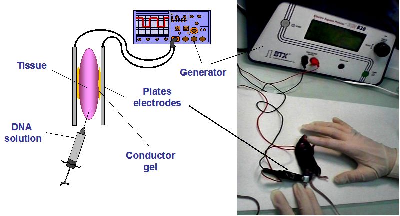
The in vivo electrotransfer technique is particularly easy to implement: a solution of plasmid DNA (i. e. a circular nucleic acid) in isotonic saline (NaCl, 150mM) is injected into the target tissue with a syringe, and electric pulses are then delivered by means of electrodes placed on either side of the injection site and connected to a generator (Figure 2). Electrodes can be either needles or plates.
image4_w.jpg

Figure 2.

Experimental set up for intramuscular plasmid electrotransfer in mice

This technique allows a site specific gene transfer. It is relatively efficient in skeletal muscle and is applicable to many other tissues such as brain, liver, skin, bladder, kidney, lung, cornea, retina, testis, tumor tissue etc... for more details see [46]. Electrotransfer can also be used in a wealth of animal models, ranging from rats and mice to sheeps [47] and cows [48] and even fish [49].
5.1. Operating parameters
The efficiency of gene transfer depends on the target tissue, the delivered DNA and electric pulses parameters. The aim is to deliver, into each tissue, electrical pulses that can cause the permeabilization of cell membranes and DNA transfer, while remaining below the toxic threshold. Otherwise, local cell death by necrosis of the treated cells would occur, followed by tissue regeneration, which would induce the loss of the benefit of the treatment (but with no toxicity at the level of the whole organism). Therefore, optimal conditions for the DNA electrotransfer in a targeted tissue result from a compromise between the efficiency of DNA transfer and minimal cellular toxicity.
5.1.1. The electrodes
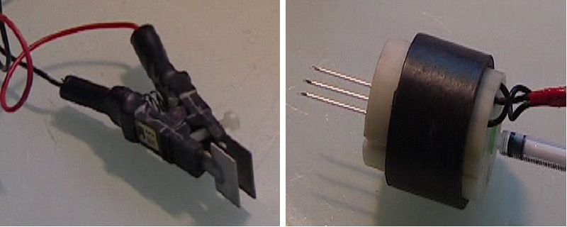
The choice of electrodes depends on the target tissue and the size of the treated animal. It is critically important and should be carefully considered. For an electrotransfer on a small animal in a tissue such as skeletal muscle, or liver tumor, most experimenters use electrodes made of two plates attached to a clamp (Figure 3, left). Indeed, this type of electrodes can be easily applied externally on each side of the interested tissue. Because one key parameter is the electric field, which is related to the ratio between the voltage applied and the distance between electrodes, this latter distance should not be too large in order to avoid prohibitive high voltage in vivo delivery. Thus, for animals of larger size, needle electrodes (Figure 3, right) are more often used than external plates.
image5_w.jpg

Figure 3.

Examples of electrode plates for external use (left) and needle electrodes for internal use, designed by the company Sphergen (right).

5.1.2. Electrical parameters
Knowing the magnitude and distribution of electric field is very important for both efficient gene transfer and reduced toxicity. The distribution of the electric field is dependent on both the tissue and the type of electrodes, which causes variations in the effective magnitude of the field in the tissue area of interest. The electric field distribution is more homogeneous when using plate electrodes than with needle electrodes, and for a given setting, the resulting electric field is lower with needle electrodes that with electrode plates [38].
Moreover, it is necessary to determine, for each tissue and each species, the threshold values of the electric field magnitude, i.e. the permeabilization threshold (reversible) and the cell damage threshold (irreversible), in order to define optimal electrical conditions for gene transfer with minimal toxic effects. Micklavic et al. have developed a system combining numerical predictions and experimental observations in order to determine these thresholds in the case of needle electrodes used in rat liver for drug delivery [50].
Different types of electrical pulses can be applied: unipolar square pulses, bipolar square pulses, or pulses with exponential decay [51]. The exponentially decaying pulses, colloquially referred to as “exponential pulses” are mainly used in vitro with a time constant dependent on the resistance of the incubation media. The square pulses are preferred in vivo, since the voltage and pulse duration can be set independently of the electrical resistance of the targeted tissue. Unipolar square pulses are the most widely used for electrotransfer experiments, while bipolar squares pulses are rather used for electrophysiology [52].
5.2. Toxicity
Tissue damage can be caused by electrotransfer and thus limits the efficiency of transfection [53]. The cell permeabilization is the main toxicity factor: it leads to an inward diffusion of the external medium as well as leakage of intracellular content, thus changing the composition of the latter. This toxicity can be reduced by minimizing the duration and the extent of permeabilization.
Other factors of toxicity have been described such as oxidative stress due to the formation of free radicals near the electropermeabilized membrane [6, 54]. It was also shown that electrotransfer induced muscle damage dependent on the amount of DNA injected [55]; these lesions disappear within two months after injection.
In our laboratory, histological analyzes of muscle slices have shown that the application of electric fields optimized for gene transfer does not induce gene expression markers of stress and cellular toxicity [56]. Other experiments have allowed to conclude that, even in optimized conditions, very little muscle damage is generated: few inflammatory lesions are observed with a maximum in the first seven days after the electrotransfer, but these disappear rapidly in less than three weeks [57-58].

 
Last edited:

China Connection

TB Fanatic
It is also possible to reduce the extent of damage by increasing the accessibility of DNA to target cells. Indeed, studies have shown that improving the plasmid distribution leads to an increase in transgene expression. Thus, the value of the electric field used can be reduced. Better distribution can be obtained for example by pre-injection of hyaluronidase [59], an enzyme that degrades hyaluronic acid, which is a major component of the extracellular matrix [60]. This pretreatment allows for the same expression level, using lower voltages while reducing muscle damage [61]. A pre-injection of sucrose may also improve the distribution of DNA, by creating spaces between the muscle fibers [62]. Similarly, a pre-injection of poly-L-glutamate, an anionic polymer, seems to increase the internalization of the plasmid inside the cell and/or to reduce its degradation [63], and therefore increases the expression of exogenous gene.
5.3. Target tissues
During recent years, electrotransfer has been applied in various animal species to many tissues, including skeletal muscle, skin, liver, lungs, kidneys, joints, brain, retina, cornea, etc... [64]. The optimal parameters of a given electrotransfer should be determined based on the cell type and species, since these parameters strongly depend on tissue organization and the size of the transfected cells.
5.3.1. Skeletal muscle
One of the most widely used tissues for electrotransfer is skeletal muscle. The DNA electrotransfer into skeletal muscle was discovered independently by three teams [8, 9, 52]. Indeed, skeletal muscle offers many advantages:
  • a large, easy access;
  • sets of muscle fibers are parallel to each other: many fibers might have an optimal orientation relative to the electric field, which promotes even transfer across the entire length of the fibers;
  • unlike other cells, muscle cells have multiple nuclei flattened against the cell membrane, which facilitates DNA trafficking to the nucleus;
  • muscle fibers do not divide, ensuring long-term gene expression, notwithstanding the absence of regeneration due to injury or cytotoxic immune response;
  • finally, a major advantage of skeletal muscle lies in its ability to produce and release biologically active proteins into the bloodstream, due to the strong vascularisation.
Combined together, these features can turn muscle into systemic drug delivery system for distant targets [65]. Interestingly, the cotransfection of multiple unlinked genes can be easily performed by electroporation [66]. For examples of electrotransfer in skeletal muscle in various mammalian species see [46].
5.3.2. The skin
The skin is, as muscle, also a widely used tissue for DNA electrotransfer, mostly because:
  • this tissue is easily accessible and a large area of tissue can be treated;
  • keratinocytes, which are epidermal cells, can synthesize and secrete therapeutic proteins that reach the bloodstream;
  • by its natural function of a biological barrier, the skin contains cells that present antigens and is therefore an organ of choice for applications in DNA vaccination;
  • the epidermal cells have a short lifespan, which can be useful for treatments requiring a brief period of expression.
However, skin structure [67] does not facilitate gene transfer. In particular, the top layer (stratum corneum or horny layer) is a major barrier [68, 69]. But a high level of expression in the skin from a single injection could still be observed [70, 71]. Moreover Dujardin et al. have shown that square or exponential pulses induce moderate and reversible effects on the skin without inflammation or necrosis, while transiently permeabilizing the skin and thus allowing the passage of molecules [72].
5.4. Optimization of in vivo electrotransfer conditions
An important goal for gene transfer applications is the level and duration of gene expression. To determine optimal conditions which maximize efficiency while reducing tissue damage, different protocols have been used to improve the access of plasmids to targeted cells. As already described, improved plasmid distribution in the skeletal muscle leads to an increase in DNA expression. Accordingly, Cemazar et al. showed recently enhanced transfection efficiency of gene transfer by pretreatment of tumors with hyaluronidase and/or collagenase, two enzymes which modulate components of the extracellular matrix [73].
A secretion signal can be also added to the transgene sequence : we have recently shown that by modifying the cellular localization of the produced protein by adding a secretory signal, the production and secretion of this protein is enhanced, thus enhancing biological effect [74].
We have also shown that codon optimization of the transgene (i.e. retaining the natural amino acid sequence but using the preferred host animal codons) leads to increase in the expression of the protein of interest [74].
Another method to increase the stability of the protein produced in the blood circulation is to increase its size in order to avoid kidney excretion. Thus, the construction of fusion proteins, for instance by fusing a therapeutic protein with an IgG constant [75], appears a simple way to deliver enhanced levels of secreted proteins without altering their biological activities.
The enhanced protein expression, and so their biological effects, also depends of the injection regimen and the administered plasmid dose [74].

6. Applications of plasmid electrotransfer
DNA electrotransfer is a recent technique of has not yet successfully completed all stages of clinical development, but this is progressing. The first Phase I human clinical trial has been initiated in U.S. by the company Inovio Biomedicals, for the treatment of skin cancer [76]. Since then, the delivery of plasmid DNA encoding therapeutic genes has been tested extensively in preclinical melanoma models [77].
Applications designated as "therapeutic" which are mainly reported in the literature have been demonstrated on animal models of human diseases. The main potential therapeutic areas cover cancer [78], cardiovascular diseases [75], autoimmune diseases [79], monogenic diseases [80], organ-specific disorders [81] and vaccination [82, 83]. Different examples show the efficiency of plasmid electrotransfer to produce therapeutic proteins in various pathologies [46]: all these experiments showed an improvement in symptoms of the relative disorder.
6.1. Cancer
Cancer accounts for major field of application trials of gene therapy. Different strategies can be broadly grouped into four main categories:
  1. Stimulation of the immune response against a tumor [84],
  2. Use of suicide genes [85-87];
  3. Repair cell cycle defects caused by the loss of tumor suppressor genes or oncogene activation [88],
  4. Inhibition of tumor angiogenesis [89].
These strategies can be combined to obtain synergistic results. For example, a combination of HSV-TK-suicide gene therapy and IL-21 immune gene therapy byelectrotransfer improves antitumor responses in mice [90]. Moreover, in vivo electrotransfer could be used in combination with other strategies such as chemotherapy, because these two approaches use different mechanisms to kill cancer cells, and thus a synergistic effect may be obtained.
Actually, electroporation of DNA encoding cytokines into tumors is extensively studied: IL-12 [91], IL-18 [92], IFN-α [93] have been shown to reduce tumor growth and increase survival times in different tumor models. Other interesting results are represented by the inhibition of tumor growth in various models with plasmids encoding metaloproteinase-3 inhibitor for the treatment of prostate cancers [94], or encoding endostatin for his therapeutic efficacy in mouse-transplanted tumors [95].
All these experiments show the potential of in vivo electrotranfer for cancer treatment. And the strategy used, i.e. the direct intra-tumoral plasmid electrotransfer, is well suited for the local production of therapeutic proteins. However, since the efficacy of gene transfer into tumor cells in vivo is generally low, intramuscular electrotransfer can also be efficiently used for distal tumor treatment. Indeed, an important application of the technique of plasmid electrotransfer is the protein secretion by skeletal muscle: the produced protein, such as, for instant, an immunostimulating cytokine, can diffuse into the vascular system and circulate throughout the body to exert a physiological effect, particularly therapeutic. This distal approach may be very powerful for surgically inaccessible tumors, such as head and neck tumors.
Finally, the intramuscular electrotransfer of a plasmid encoding the prostate membrane specific antigen (PMSA) has been tested in a human clinical trial of prostate cancer active immunotherapy [96]. DNA fusion-gene vaccination in patients with prostate cancer induces high-frequency CD8 (+) T-cell responses and increases PSA doubling time [97].
 

China Connection

TB Fanatic
6.2. Monogenic diseases
Monogenic diseases with an identified defective gene have been the first diseases targeted by gene therapy approaches. Among these diseases, Duchenne muscular dystrophy (DMD), which is characterized by the absence of dystrophin, is a good model, since even a small amount of dystrophin would be sufficient to reverse the clinical phenotype of the disease. An approach to eventually restore this protein in patients with DMD is to introduce into their muscles a plasmid encoding dystrophinc DNA. Pichavant et al. were the first to demonstrate local restoration of full-length dog dystrophin in dystrophic dog muscle by DNA electrotransfer [98].
6.3. Hematopoietic factor deficiency
Erythropoietin (EPO) is another good candidate for gene therapy applications because a small amount will produce the desired physiological effect of raising the hematocrit. Numerous studies, in particular by our own group, report efficient EPO secretion after plasmid electrotransfer, with a therapeutic effect in anemia and beta thalassemia. The use of intramuscular plasmid electrotransfer for EPO gene delivery in mice increased approximately 10 to 100-fold the expression of this gene, as compared to naked DNA alone [99, 100]. Moreover with this method, the protein in circulation and hematocrit levels were stable for 2 to 6 months after a single injection of minimal amounts (as little as 1 µg) of a plasmid carrying the mouse EPO cDNA. Several studies also showed that EPO expression could be regulated, for instance by co-administering an EPO encoding plasmid under the control of a tetracycline-inducible promotor and a second plasmid carrying the reverse tetracycline-dependent transactivator protein [100, 101]. All these studies exemplified that plasmid DNA electrotransfer can efficiently produce enough amounts of transgenic EPO in normal mice.
In collaboration with the group of Y. Beuzard, we have demonstrated the relevance of intramuscular electroporation of an EPO-expressing plasmid in a mouse model of human β-thalassemia, a severe genetic disease, leading to a durable and dose-dependent phenotypic correction of this severe genetic disease [102]. In addition, we have also shown that it is possible to produce fusion protein by plasmid DNA electrotransfer [103]: indeed since the bridging of two adjacent EPO receptors triggers a conformational change that initiates signal transduction [104], we have hypothesized that the fusion of two EPO molecules might lead an increase in intrinsic activity of EPO. Thus, we demonstrated that the injection of EPO dimer encoding plasmid by electrotransfer in a skeletal muscle of β-thalassemic mice induces an increase in the biologic specific activity of this EPO dimer in comparison with the activity of monomer [103].
Furthermore the secretion peak of therapeutic protein following DNA administration is potentially deleterious. We reported that muscular electrotransfer of low doses of plasmid can be repeated several times to weeks or even months after the initial injection, and that this strategy leads to efficient, long-lasting and non-toxic treatment of β-thalassemic mouse anemia avoiding the deleterious initial hematocrit peak and maintaining a normal hematocrit with small fluctuation [105].
In addition, Gothelf et al. demonstrate that gene electrotransfer to skin of even small amounts of EPO DNA can lead to systemically therapeutic levels of EPO protein [106].
6.4. Cardiovascular diseases
Gene therapy is an attractive strategy for the treatment of cardiovascular disease. However, using current methods, the induction of gene expression at therapeutic levels is often inefficient. Therefore DNA electrotransfer directly into heart may enhance the delivery of therapeutic protein as shown the team of R. Heller : the electroporation method ameliorates the delivery of a plasmid encoding an angiogenic growth factor (vascular endothelial growth factor, VEGF), which is a molecule previously documented to stimulate revascularization in coronary artery disease [107]. Ayuni et al. demonstrated that, unlike the usual methods to treat coronary artery diseases, electrotransfer applied directly into the beating heart enhances the delivery of a plasmid injected via the coronary veins after transient occlusion of the coronary sinus [108]. These results show that in vivo electroporation mediated gene transfer is feasible and safe,in particular to the heart. Finally, in skin, D. Dean reported that using electroporation in skin enhances delivery of plasmid DNA encoding fibroblast growth factor-2 (FGF-2) to induce neovascularization as atherapy for ischemia in a rat model [109].
6.5. Eye diseases
The eye is an isolated organ difficult to reach via systemic administration. Eye diseases are treated with intra- or periocular injections and these repeated injections bear the risk of adverse effects, mainly infections, and are poorly tolerated by the patients. The use of DNA electrotransfer technique is therefore possible to deliver a local treatment. Our team associated with an ophtalmology group has developed electrotransfer to the ciliary muscle, which is a particular smooth muscle with some characteristics of striated skeletal muscle, for the local treatment of inflammatory eye disease. This approach led to production and secretion of therapeutic levels of TNFα soluble receptor in the ocular media, and not in the serum, thus preventing clinical and histological signs in a rat uveitis model [110, 111]. Recently, suprachoroidal electrotransfer with a reporter plasmid to transfect the choroid and the retina without detaching the retina has been reported [112]. Not only choroidal cells but also RPE, and potentially photoreceptors, were efficiently transduced for at least a month, without ocular complications. This minimally invasive non-viral gene therapy method may open new prospects for human retinal therapies.
6.6. Obesity and diabetes
As mentioned above, skeletal muscle can be an efficient platform for the secretion of erythropoietin (EPO), which displays a variety of metabolic effects when it is expressed in supra-physiological levels. Hojman et al. have proposed to overexpress EPO in muscle by electrotransfer of plasmid in the aim to protect mice against diet-induced obesity and normalize glucose sensitivity, associated with a shift to increased fat metabolism in the muscles [113]. Similar results were obtained after DNA electrotransfer of plasmid encoding the carnitine palmitoyltransferase 1 (CPT1), the enzyme that controls the entry of long-chain fatty acyl CoA into mitochondria: an overexpression of CPT1 led to enhance rates of fatty acid beta-oxidation and improved insulin action in muscle in high-fat diet insulin-resistant rats [114]. In the same model, electrotransfer of the orphan nuclear receptor (Nur77) significantly ameliorates the effect of this protein on glucose metabolism [115].
6.7. Vaccination and passive immunization by antibody production
The prospect of inducing an immune response to a protein expressed in vivo directly from administered DNA vaccine represents an attractive alternative to other modes of vaccination. Plasmid electrotransfer has been used in genetic immunization to produce antigenic proteins. It is now well established that genetic immunization induces both durable cellular and humoral responses [116]. This type of immunization is often developed for vaccination (virus or antibacterial), for anticancer active immunotherapy, and also to induce in animals the production with high yield of antibodies against a given antigen.
Since electrotransfer efficiently transfers genes compared to a single injection of plasmid, improving antigenic protein expression by several orders of magnitude, the antibody titer and the quality of the immune response are also improved [117], with an increasing factor of 100 in mice after electrotransfer of a plasmid encoding a surface antigen of hepatitis B [118]. High titers of antibodies were also obtained in mice and rabbits after i.m. electrotransfer of a plasmid encoding an envelope glycoprotein of hepatitis C [119], and in mice after electrotransfer of a plasmid encoding a protein of Mycobacterium tuberculosis [120]. In the laboratory, it was shown that i.m. electrotransfer of a plasmid encoding the influenza hemagglutinin induces a better immune response in mice that a single i.m. injection [121]. And recently, we have assessed the potential of i.m. electrotransferin mouse to produce neutralizing antibodies, with high titer, against botulinum toxins, the most powerful poison in the world in present time [74]. We have optimized DNA electrotransfer for genetic immunization against botulinum antigen. This DNA immunization has been used in rabbits to induce antibodies production which is compatible with industrial development of antiserum production for a human therapeutic use (Burgain et al., unpublished results).These examples show that it is possible to obtain high titers neutralizing antibodies in animals by DNA electrotransfer.
Monoclonal antibodies are increasingly being used in a wide range of clinical applications in the field of autoimmune disease, cancer and infectious disease. The production and secretion by electrotransfered muscle of monoclonal antibodies has been demonstrated by our group and the one of I. Mathiesen, independently [83, 122]. These studies demonstrated that the co-transfection of two naked plasmids encoding the heavy and light G immunoglobulin chains led to the secretion of fully assembled and functional immunoglobulin molecules. The successful neutralization of various pathogens resulted from monoclonal antibody secretion by electrotransfered muscle, raising the possibility of clinical passive immunization applications.
7. Conclusion
In vivo electrotransfer is a non-viral technique which has emerged as an efficient, user-friendly and cheap gene transfer method which issuited for a wide range of tissues and species. Moreover, in vivo electrotransfer can be used for either local or distal effect by secretion of the transgenic protein into the bloodstream. The skeletal muscle is able to produce functional proteins with adequate post-translational modifications, which means that the muscle can be used as an endocrine organ for the production of therapeutic secreted proteins targeting systemic diseases. It is now established that therapeutic levels of circulating proteins can be reached in animal models. And since DNA does not induce any immune response, plasmid electrotransfer can be repeated as often as desired (Scherman et al., unpublished results).
The understanding of the precise mechanism of electrotransfer, the optimization of its realization, the improvement of plasmids and of the structure of the encoded protein will bring more efficiency and above all more safety to the method, should it be applied to humans. Several clinical trials have been conducted and/or are still in progress. For more details see Search of: electroporation - List Results - ClinicalTrials.gov. These clinical trials are mainly conducted against infectious diseases such AIDS, hepatitis B, malaria, dengue, influenza... and various cancer types such as ovarian cancer or renal cancer, melanoma, cancers caused by human papillomavirus... Thus, DNA electrotransfer appears as a powerful and promising tool not only for gene therapy, but also for in vivo gene delivery at the laboratory level within the frame of physiological studies.

 
Top